

Pregnancy
Congratulations on your pregnancy!
Pregnancy is a very special phase of life - a phase of upheaval, of physical and emotional changes, of anticipation and longing expectations. But it is also a time of uncertainty, questions and fears. That is normal and understandable.
We are happy to be able to support our pregnant patients during these important weeks and months and thus help them to enjoy their pregnancy without any stress.
The focus of our practice is the support and care of expectant mothers in accordance with the maternity guidelines as well as supplementary, comprehensive prenatal ultrasound diagnostics (Degum II). The ultrasound examinations are performedwith a modern ultrasound machine with high resolution and 3D/4D technology. All our pregnant patients have the option of accessing the ultrasound images via a personal user account at FetView, viewing them at home on a PC, smartphone or tablet PC, saving them, copying them or sending them by e-mail.
Regular check-ups are important for the health of the child and the well-being of the mother. All examinations that are part of antenatal care according to maternity guidelines are covered by the statutory health insurance funds. In order to provide our patients with optimal maternity care with comprehensive prenatal diagnostics beyond what is covered by the statutory health insurance, we offer the following examinations:
Prenatal diagnostics
Prenatal DNA test, trisomy blood test, Down's syndrome blood test
Implementation: from 11. SSW
The prenatal test is used to determine the risk of chromosomal disorders, the so-called trisomies. The examination is non-invasive, i.e. it is not associated with any risks for the child or the pregnant woman. A simple blood sample from the expectant mother is sufficient to obtain the material needed for the test.
The prenatal blood test has a high detection rate, but does not provide a one hundred percent reliable diagnosis. This is only possible with a chorionic villus sampling (tissue sampling from the placenta, from the 11th week of pregnancy) or an amniocentesis (amniocentesis, from the 15th week of pregnancy). With the blood test, however, these invasive examinations, which are associated with an intervention-related risk of miscarriage of approx. 0.5%, can be kept to a minimum.
What is being examined?
The prenatal blood test determines the risk for trisomy 21 (Down's syndrome), trisomy 18 (Edwards' syndrome) and trisomy 13 (paetau's syndrome). As trisomy 21 is the most common form of trisomy, the prenatal test is also referred to as the Down syndrome blood test.
In addition, the prenatal blood test can determine the sex chromosomes (X, Y) and the risk of anomalies such as Turner syndrome or Klinefelter syndrome.
What cannot be examined?
With the help of the blood test, only a part of the possible chromosomal disorders can be detected. For example, the test is not suitable for detecting partial trisomies, in which only part of the chromosomes are present in triplicate. Hereditary diseases such as cystic fibrosis cannot be detected either.
The test does not replace fetal ultrasound diagnostics. It does not detect any of the frequently occurring fetal malformations if they are not based on a chromosomal disorder. It is therefore only useful in combination with an early differentiated ultrasound.
How accurate is the prenatal test?
The prenatal test determines the risk for the chromosomal disorders mentioned with high accuracy and at an early stage during pregnancy. The detection rate of the Harmony prenatal test, which we use in our practice, is over 99% for trisomy 21 (comparison: first trimester screening with nuchal translucency measurement 90%).
In individual cases, the test does not provide a usable result. This is the case, for example, if the proportion of the child's DNA in the mother's blood is too low.
Procedure and costs:
The blood sample can be taken from 10+0 weeks of pregnancy after detailed genetic counselling and in combination with an early fine diagnostic ultrasound. The result of the Harmony prenatal test is generally available within 9 days. The costs for the trisomy blood test and the associated medical services have not been covered by health insurances so far. It is planned to cover the costs in risk cases and for clarifying abnormalities.
Nuchal translucency measurement, first trimester screening
Implementation: 12 - 14 SSW
A nuchal translucency measurement as part of a comprehensive first trimester screening provides you with a lot of information about your child's development and health at a very early stage. In addition to a risk assessment for Down's syndrome (trisomy 21), indications of serious organ malformations such as heart defects can be recorded.
The purpose of a nuchal translucency measurement is to make a risk assessment and to give the pregnant woman a decision-making aid if she is not yet sure whether she wants to have an amniocentesis.
Procedure of the examination
The focus of the first trimester screening is a comprehensive ultrasound examination, which can already exclude numerous organic malformations. In order to assess the risk of a chromosomal defect, the so-called nuchal translucency of the unborn child is measured by ultrasound. This structure can generally be seen in all foetuses during this examination period. However, if the NT is widened, the probability of the child having a disease increases.
In addition to the ultrasound examination, two values are determined in the maternal blood: the pregnancy hormone ß-HCG and the protein PAPP-A. The blood sample can be taken as early as 10+0 weeks gestation, i.e. about 2 weeks before the ultrasound examination.
Risk calculation
The individual risk for the presence of Down's syndrome can be calculated from the following findings:
- the age of the mother
- the exact age of pregnancy
- previous pregnancies with chromosomal defects
- the width of the nuchal translucency of the unborn child
- the values of the blood analysis
With the help of this method, which is not associated with any risks for mother and child, about 90% of affected foetuses are detected. However, the diagnosis must be confirmed by a tissue sample from the placenta(chorionic villus sampling) or anamniocentesis.
Cost absorption
The costs for a nuchal translucency measurement - like all examinations for risk assessment (e.g. the prenatal DNA test) - are not covered by the statutory health insurance funds. However, if abnormal test results lead to further examinations, these can be settled via the health insurance fund.
A request from our practice
During the first trimester screening, we concentrate entirely on you and your unborn child. Therefore, we ask you not to bring smaller siblings with you during this demanding and time-consuming examination. Thank you very much.
Fine diagnostic ultrasound / malformation diagnostics (DEGUM II)
Implementation: 20th - 22nd SSW
The organ ultrasound (also called fine diagnostics or differentiated ultrasound) is much more extensive than the ultrasound examination provided for in the maternity guidelines during this period. In addition to special equipment, it requires a special qualification(DEGUM II) of the examiner.
Procedure of the examination
Under good examination conditions, where the position of the child and the thickness of the maternal abdominal wall play a decisive role, a comprehensive fine diagnostic examination takes about 50 - 60 minutes. All the organs and features of the unborn child that can be visualised are examined and the following points are assessed:
- the age-appropriate growth of the child
- the amount of amniotic fluid and the position and appearance of the placenta
- the appearance and function of all visible organs including the heart
- The blood flow in the umbilical cord and the blood flow in the uterine vessels (to assess placental function and maturation).
 Meaning and consequences
The primary purpose of this comprehensive examination is to make an exact diagnosis in the case of abnormalities in the screening and to accompany high-risk pregnancies.
If a disease or malformation of the child is detected, we can derive consequences for the further course of pregnancy (e.g. further diagnostics or therapies) or for the birth (e.g. involvement of further specialists, choice of maternity hospital) together with the patient in good time.
For many parents-to-be, the ultrasound helps to deepen the relationship with their unborn child. An inconspicuous organ ultrasound brings you security and reassurance for the further course of the pregnancy. However, every prenatal examination can also reveal unexpected abnormalities and present the parents with the decision about further examinations and consequences.
Cost absorption
For certain indications, the costs of fetal malformation diagnostics are covered by the statutory health insurances - e.g. in the case of conspicuous previous findings or if a couple already has a sick or disabled child. Otherwise, we offer this examination as an individual health service (IGeL).
A request from our practice
During the fine diagnostic organ ultrasound, we concentrate entirely on you and your unborn child. Therefore, we ask you not to bring smaller siblings with you during this demanding and time-consuming examination. Thank you very much.
Fetal echocardiography / ultrasound examination of the fetal heart
Implementation: from 19+0 SSW
Fetal echocardiography is part of the fine diagnostic assessment of the child's organs. The sonographic examination of the fetal heart requires great experience on the part of the examiner as well as a high-resolution ultrasound machine.
Although some heart malformations can be detected during early ultrasound screening between the 13th and 14th week of pregnancy, a detailed assessment of the heart, its function and blood flows only takes place between the 20th and 22nd week of pregnancy.
Procedure of the examination
During a comprehensive fetal echocardiography, the following structures and functions are assessed:
- Position, size and symmetry of the heart
- Anatomy of the heart structures
- Function of the heart valves
- Heart beat rate and heart rhythm
- Location of the major arterial and venous vessels
Further details are examined with the help of colour-coded Doppler sonography:
- Function of the ventricles
- Cardiac septums
- Blood flows in the heart (according to their direction and speed)
- Blood flows in the major arterial and venous vessels (according to their direction and velocity).
Meaning and consequences
The majority of congenital heart defects can be corrected after birth by one or more operations. The life and health of the child depend decisively on the early and accurate diagnosis of the malformation.
If a child is diagnosed with a heart defect, we will immediately refer you to a specialist (usually a paediatrician or obstetrician specialising in cardiology) with whom you can discuss the further procedure.
Heart defects often occur in combination with chromosomal defects or genetic syndromes. Therefore, further prenatal diagnostics may be useful in the case of abnormal examination findings of the child's heart.
Extended ultrasound examinations
According to the maternity guidelines, only three ultrasound examinations are covered by health insurance during an unremarkable pregnancy.
Routine ultrasound screening is carried out at the following weeks of pregnancy:
- Between the 9th and 12th class
- the 19th and 22nd
- and the 29th and 32nd week of pregnancy
Ultrasound examinations outside these times are considered to be self-responsible health services in the case of an inconspicuous pregnancy, the costs of which are not covered by the health insurance funds.
During an extended ultrasound examination, we take more time to show the expectant parents the baby and - if it can be seen - to determine the sex. Towards the end of the pregnancy, an extended ultrasound scan can check the position of the baby to make sure that the head is in the pelvis. In addition, the placenta, the amount of amniotic fluid and the size ratio of the baby's head to the mother's pelvis can be assessed.
You can view, save, copy or send the ultrasound images via a personal user account at FetView at home on your PC, smartphone or tablet PC.
Colour Doppler ultrasound
Carried out from the 27th week of pregnancy
Doppler ultrasound can be used to visualise the blood flow in the fetal vessels and in part of the maternal vessels and to assess the supply to the unborn child. Doppler ultrasound is mainly used in late pregnancy (26 - 38 weeks) and is not associated with any risk for mother and child.
A Doppler examination is justified in the case of
- Suspicion of reduced growth or growth arrest of the child
- Decreased amniotic fluid
- Suspicion of child malformation or disease
- pregnancy-related illness of the mother (e.g. high blood pressure, pre-eclampsia, diabetes, kidney disease)
- certain infections (e.g. ringworm)
- Premature birth or birth defects in a previous pregnancy
- Multiple pregnancies
Procedure of the examination
During each Doppler examination, we first assess the baby's growth, the amount of amniotic fluid and the maturation of the placenta. Then we measure
- The blood flow in the fetal vessels (e.g. aorta, cerebral vessels, umbilical cord).
- the blood flow in the uterine vessels
Meaning and consequences
The colour Doppler ultrasound provides information about acute or chronic deficiencies in the supply of the unborn child and about the function of the placenta. On the one hand, the Doppler examination can help to reassure the parents-to-be if an initial suspicion is not confirmed. For example, if the child is too small for the gestational age, but the supply of the child is still good.
On the other hand, situations in which action is needed can be recognised at an early stage. For example, intensive prenatal care or, in individual cases, premature delivery may become necessary.
Cost absorption
Doppler sonography is not part of the normal routine check-up during maternity care. However, in the case of conspicuous preliminary findings, the costs are covered by the statutory health insurances.
3D/4D ultrasound
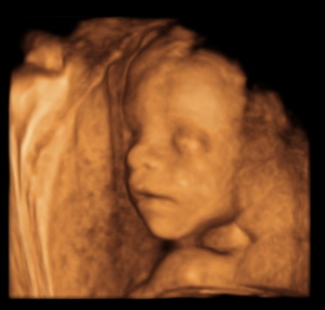
The 3D ultrasound enables a three-dimensional, spatial representation of your child or individual organs and body parts. The procedure is no different from conventional ultrasound. We speak of a 4D ultrasound when motion sequences are also documented and time is thus taken into account as the fourth dimension (e.g. through film or video recordings).
In prenatal diagnostics, the technique of 3D ultrasound is used alongside conventional two-dimensional sonography to visualise normal and abnormal fetal structures. It is mainly used when additional diagnostic indications are to be expected for special questions.
What is special about 3D ultrasound?
The 3D ultrasound does not provide a better or more accurate image due to its lower resolution, but because the three-dimensionality accommodates our visual habits, parents-to-be often find these ultrasound images particularly impressive.
For many parents, this form of ultrasound is a good way to strengthen the early bond with the unborn child. However, since every ultrasound also examines the child's development, such an examination is never a mere photo shoot. It always serves diagnostic purposes as well.
Are there always good photos?
Whether the souvenir photos from the 3D ultrasound are really successful and meet the parents' expectations depends on good viewing or sound conditions. The position of the child, a placenta lying against the anterior wall, little amniotic fluid or a thick abdominal wall often do not allow a good three-dimensional image. On average, sufficient visibility can be expected in about 50% of the examinations, depending on the week of pregnancy.
3D/4D ultrasound as a self-pay service
We offer our patients this examination as part of an extended ultrasound as a self-pay service. You can print out the images of your baby and take them home with you. With a personal user account at FetView, you also have the option of accessing your images online, viewing them at home on your PC, smartphone or tablet PC, saving them, copying them or sending them by e-mail.
More about 3D ultrasound: Frequently asked questions
Extended antenatal care / laboratory diagnostics

Our blog on the topic
FAQ
Frequently asked questions