From the desire to have children to breastfeeding
For many of us, children are the living expression of an intimate relationship and a complete family. They are an important part of our lives and so the medical support of the desire to have children, pregnancy, postpartum and breastfeeding is one of the central tasks of our practice.
The wish for a child is not always fulfilled quickly and without problems. According to estimates, about 12 - 15 % of all couples of reproductive age remain involuntarily childless. A couple is considered involuntarily childless if no pregnancy has occurred after one year of unprotected intercourse.
Women as well as men experience it as very painful that the longed-for offspring does not materialise. We refer couples who want to have children to specialised colleagues. We can accompany hormonal infertility treatment with regular follicle measurement.
An ultrasound examination can examine the structure of the uterine lining and measure the size of the follicle (egg sac). By measuring the follicle, the exact time of ovulation and thus the most favourable time for fertilisation can be determined.
Generally, follicle measurement is carried out between the 11th and 13th day of the cycle - shortly before ovulation, which is expected between the 14th and 16th day of the cycle in a regular 28-day cycle.
Website of the Federal Centre for Health (BZgA) on the topic of wanting children
Pregnant? Congratulations!
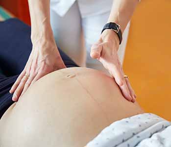
Pregnancy is a very special phase of life - a phase of upheaval, of physical and emotional changes, of anticipation and longing expectations. But it is also a time of uncertainty, questions and fears. That is normal and understandable. We are happy to be able to support our pregnant patients during these important weeks and months and thus help them to enjoy their pregnancy without any stress.
The most important focus of our practice is the support and care of expectant mothers in accordance with the Maternity guidelines as well as supplementary, comprehensive prenatal ultrasound diagnostics (Degum II). The ultrasound examinations are performed with a modern ultrasound machine with high resolution and 3D/4D technology.
Regular check-ups are important for the health of the child and the well-being of the mother. All examinations that are part of antenatal care according to maternity guidelines are covered by statutory health insurance.
From the detection of pregnancy until birth:
Regular checks of blood, urine, blood pressure, weight and - from the 28th week of pregnancy - the baby's heart tones.
9 - 12 SSW:
Your maternity record will be issued. First basic ultrasound examinationBlood tests (blood group, antibodies, rubella titer, syphilis, hepatitis B, HIV if necessary), chlamydia test.
19 - 22 SSW:
second basic ultrasound examination / extended basic ultrasound
Rhesus NIPTDetermination of the fetal rhesus factor from the maternal blood (in rhesus factor-negative pregnant women).
from 25 weeks gestation:
Simple diabetes screening, so-called small sugar test
29. – 32. SSW:
Aufzeichnung der kindlichen Herztöne (CTG), dritte Basis-Ultraschalluntersuchung, Kontrolle der Antikörper im Blut.
Anti-D-Prophylaxe: Patientinnen, die das Blutgruppenmerkmal Rhesus-negativ tragen, erhalten eine Spritze (Anti-D), um eine mögliche Erkrankung des Kindes im Mutterleib zu vermeiden.
Download flyer
Taking medication during pregnancy and breastfeeding
Website of the Charité in Berlin on drug safety during pregnancy and lactation
with search function for drugs and active ingredients
Website of the Federal Centre for Health Education (BZgA)
with detailed information for expectant parents and valuable tips for pregnancyspezielle Informationen zum Corona-Virus und einer Infektion während der Schwangerschaft
Prenatal diagnostics
In order to provide our patients with optimal maternity care with comprehensive prenatal diagnostics, we offer the following examinations as self-administered health services (IGeL):
Implementation: 12 - 14 SSW
Comprehensive first trimester screening with a detailed organ ultrasound and nuchal translucency measurement gives you a lot of information about your child's development and health at a very early stage. In addition to a risk assessment for the Down syndrome (trisomy 21) further chromosomal defects and serious organ malformations, such as heart defects, can be detected. First trimester screening with an early organ ultrasound is also recommended for young pregnant women. The general risk for malformations of 3 - 5% is independent of age
Which examinations are part of the first trimester screening?
1. early organ ultrasound
First trimester screening focuses on a comprehensive ultrasound examination, which can be performed between 11+1 and 13+6 SSW. The ideal examination period is between 12+0 and 13+6 SSW. Under good examination conditions, we can go into details of the child's development and organs and already exclude numerous organic malformations. However, a strong maternal abdominal wall or an unfavorable position of the child may limit the examination possibilities.
2. nuchal translucency measurement
In order to assess the risk for the presence of a chromosomal disorder, the so-called nuchal translucency of the unborn child (also called nuchal fold or NT for "nuchal translucency") is measured during the ultrasound examination. This structure can basically be seen in all fetuses during the mentioned examination period. However, if the NT is widened, the probability of the child having a disease (e.g. Down syndrome and/or heart defect) increases. The measurement requires a good resolution ultrasound scanner and experience of the examiner.
About two weeks before the ultrasound examination, blood will be taken from you as part of the first trimester screening. Two different blood tests can be performed: The non-invasive prenatal DNA test (NIPT, Harmony test) or the classic blood test to calculate the risk of trisomy. However, both tests are only useful in combination with early organ ultrasound.
Cost absorption
The costs for the detailed first trimester screening with early organ ultrasound and nuchal translucency measurement are not covered by the statutory health insurance funds.
A request from our practice
During the first trimester screening, we focus entirely on you and your unborn child. Therefore, we ask you not to bring younger siblings with you during this demanding and time-consuming examination. Thank you very much.
Implementation: from 11. SSW
The prenatal test is used to determine the risk of chromosomal defects, the so-called Trisomies. The test is non-invasive, i.e. it is not associated with any risks for the child or the pregnant woman. A simple blood sample from the expectant mother is sufficient to obtain the material necessary for the test.
The non-invasive prenatal test (NIPT) has a high detection rate, but does not provide a 100% reliable diagnosis. This can only be achieved by Chorionic villus sampling (tissue sampling from the placenta, from the 11th SSW) or a Amniocentesis (amniocentesis, from the 15th SSW). However, the blood test can minimize these invasive examinations, which are associated with an intervention-related risk of miscarriage of up to 0.4%.
What is tested?
The prenatal blood test determines the risk for the Trisomy 21 (Down syndrome) , Trisomy 18 (Edwards syndrome) and Trisomy 13 (Pätau syndrome) . Since trisomy 21 is the most common form of trisomy, the prenatal test is also called the Down syndrome blood test.
In addition, the prenatal blood test can determine the sex chromosomes (X, Y) and determine the risk for anomalies such as Turner syndrome or Klinefelter syndrome can be determined. However, this special analysis is only recommended if there are risk factors for this (for example, anamnestic risks or abnormalities in the ultrasound).
What cannot be examined?
With the help of the blood test, only a part of the possible chromosomal disorders can be detected. For example, the test is not suitable for detecting partial trisomies, in which only part of the chromosomes are present in triplicate. It is also not possible to detect hereditary diseases such as cystic fibrosis.
How accurate is the prenatal test?
The prenatal test determines the risk for the above chromosomal disorders with high accuracy and at an early stage during pregnancy. The detection rate of the Harmony prenatal test, which we use in our practice, is over 99% for trisomy 21 (comparison: first trimester screening with nuchal translucency measurement 90%).
In individual cases, the test does not provide a usable result. This is the case, for example, if the proportion of fetal DNA in the maternal blood is too low, such as in overweight patients.
Procedure and costs
The blood sample can be taken from 10+0 weeks of pregnancy after detailed consultation. The result of the Harmony prenatal test is generally available within 5 - 10 days. Since 1.7.2022, the costs for the trisomy blood test (NIPT - non-invasive prenatal test) are covered by the health insurances if there is a medical indication (e.g. an abnormal ultrasound finding).
As an alternative to the prenatal DNA test, a blood test can be performed between the 10+0 and 13+6 SSW, in which two values are determined in the maternal blood: the pregnancy hormone ß-HCG and the protein PAPP-A.
Risk calculation
The individual risk for the presence of Down syndrome can be calculated from the following findings:
- the age of the mother
- the exact age of pregnancy
- previous pregnancies with chromosomal defects
- the width of the nuchal translucency of the unborn child
- the values of the blood analysis
With the help of this method, which is not associated with any risks for mother and child, about 90% of affected fetuses are detected. However, the diagnosis must be made by tissue sampling from the placenta (Chorionic villus sampling) or an amniocentesis (Amniocentesis).
Cost absorption
The costs of the classic blood test for risk assessment are not covered by the statutory health insurance funds. However, if conspicuous test results lead to further examinations, these can be settled via the health insurance.
Implementation: 20th - 22nd SSW
The organ ultrasound (also called fine diagnostics or differentiated ultrasound) is much more extensive than the ultrasound examination provided for in the maternity guidelines during this period. In addition to special equipment, it requires special qualifications (DEGUM II) of the examiner.
Procedure of the examination
Under good examination conditions, where the position of the child, the placenta and the thickness of the maternal abdominal wall play a decisive role, a comprehensive fine diagnostic examination takes about 30 - 45 minutes. All representable organs and features of the unborn child are viewed and the following points are assessed:
- the age-appropriate growth of the child
- the amount of amniotic fluid and the position and appearance of the placenta
- the appearance and function of all visible organs including the heart
- the blood flow in the umbilical cord and the blood flow in the uterine vessels (to assess placental function and maturation)
The primary purpose of this comprehensive examination is to make an accurate diagnosis in the event of abnormalities in the screening process and to monitor high-risk pregnancies.
If a disease or malformation of the child is detected, we can derive consequences for the further course of pregnancy (e.g. further diagnostics or therapies) or for the birth (e.g. involvement of further specialists, choice of maternity hospital) together with the patient in good time.
For many parents-to-be, the ultrasound helps to deepen the relationship with their unborn child. An inconspicuous organ ultrasound brings you security and reassurance for the further course of the pregnancy. However, every prenatal examination can also reveal unexpected abnormalities and present the parents with the decision about further examinations and consequences.
Cost absorption
For certain indications, the costs of fetal malformation diagnostics are covered by statutory health insurance - e.g. in the case of conspicuous previous findings or if a couple already has a sick or disabled child. Otherwise, we offer this examination as an individual health service (IGeL).
A request from our practice
During the fine diagnostic organ ultrasound, we concentrate entirely on you and your unborn child. Therefore, we ask you not to bring younger siblings with you during this demanding and time-consuming examination. Thank you very much.
Implementation: from 19+0 SSW
Fetal echocardiography is part of the fine diagnostic assessment of the child's organs. The sonographic examination of the fetal heart requires great experience on the part of the examiner as well as a high-resolution ultrasound machine.
Although some heart malformations can be detected during early ultrasound screening between the 13th and 14th week of pregnancy, a detailed assessment of the heart, its function and blood flows only takes place between the 20th and 22nd week of pregnancy.
Procedure of the examination
During a comprehensive fetal echocardiogram, the following structures and functions are assessed:
- Position, size and symmetry of the heart
- Anatomy of the heart structures
- Function of the heart valves
- Heart beat rate and heart rhythm
- Location of the major arterial and venous vessels
Further details are examined with the help of colour-coded Doppler sonography:
- Function of the ventricles
- Cardiac septums
- Blood flows in the heart (according to their direction and speed)
- Blood flows in the major arterial and venous vessels (according to their direction and velocity).
Significance and consequences
A large proportion of congenital heart defects can be corrected after birth by one or more operations. The life and health of the child depend crucially on the early and accurate diagnosis of the malformation.
If a child is diagnosed with a heart defect, we will immediately refer you to a specialist (usually a paediatrician or obstetrician specialising in cardiology) with whom you can discuss the further procedure.
Heart defects often occur in combination with chromosomal defects or genetic syndromes. Therefore, further prenatal diagnostics may be useful in the case of abnormal examination findings of the child's heart.
According to the maternity guidelines, only three ultrasound examinations are covered by health insurance during an unremarkable pregnancy.
Routine ultrasound screening is carried out at the following weeks of pregnancy:
- Between the 9th and 12th class
- the 19th and 22nd
- and the 29th and 32nd week of pregnancy
Ultrasound examinations outside these times are considered to be self-responsible health services in the case of an inconspicuous pregnancy, the costs of which are not covered by the health insurance funds.
During an extended ultrasound examination, we take more time to show the expectant parents the baby and - if it can be seen - to determine the sex. Towards the end of pregnancy, an extended ultrasound scan can check the position of the baby to make sure the head is in the pelvis. In addition, the placenta, the amount of amniotic fluid, and the size relationship of the baby's head to the mother's pelvis can be assessed. If sound and visibility conditions are good, 3D/4D images are also possible.
You can view, save, copy or send the ultrasound images via a personal user account at FetView at home on your PC, smartphone or tablet PC.
Doppler ultrasound can be used to visualise the blood flow in the fetal vessels and in part of the maternal vessels and to assess the supply to the unborn child. Doppler ultrasound is mainly used in late pregnancy (26 - 38 weeks) and is not associated with any risk for mother and child.
A Doppler examination is justified in the case of
- Suspicion of reduced growth or growth arrest of the child
- Decreased amniotic fluid
- Suspicion of child malformation or disease
- pregnancy-related illness of the mother (e.g. high blood pressure, Pre-eclampsia, Gestational diabeteskidney disease)
- certain infections (e.g. ringworm)
- Premature birth or birth defects in a previous pregnancy
- Multiple pregnancies
Procedure of the examination
During each Doppler examination, we first assess fetal growth, amniotic fluid volume, and placental maturation. We then measure
- The blood flow in the fetal vessels (e.g. aorta, cerebral vessels, umbilical cord).
- the blood flow in the uterine vessels
Significance and consequences
Color Doppler ultrasound provides information about acute or chronic deficiencies in the supply of the unborn child and about the function of the placenta. On the one hand, the Doppler examination can help to reassure the expectant parents if an initial suspicion is not confirmed. For example, if the child is too small for the gestational age, but the supply of the child is still good.
On the other hand, situations in which action is needed can be recognised at an early stage. For example, intensive prenatal care or, in individual cases, premature delivery may become necessary.
Cost absorption
Doppler sonography is not part of the normal routine check-up during maternity care. However, in the case of conspicuous preliminary findings, the costs are covered by the statutory health insurance.
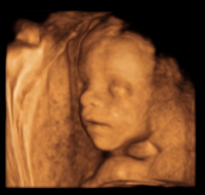
The 3D ultrasound enables a three-dimensional, spatial representation of your child or individual organs and body parts. The procedure is no different from conventional ultrasound. We speak of a 4D ultrasound when motion sequences are also documented and time is thus taken into account as the fourth dimension (e.g. through film or video recordings).
In prenatal diagnostics, the technique of 3D ultrasound is used alongside conventional two-dimensional sonography to visualise normal and abnormal fetal structures. It is mainly used when additional diagnostic indications are to be expected for special questions.
What is special about 3D ultrasound?
3D ultrasound does not provide a better or more accurate image due to its lower resolution, but because the three-dimensionality accommodates our visual habits, parents-to-be often find these ultrasound images particularly impressive.
For many parents, this form of ultrasound is a good way to strengthen the early bond with the unborn child. However, since every ultrasound also examines the child's development, such an examination is never a mere photo shoot. It always serves diagnostic purposes as well.
Are there always good photos?
Whether the souvenir photos from the 3D ultrasound are really successful and meet the parents' expectations depends on good viewing or sound conditions. The position of the child, a placenta lying against the anterior wall, little amniotic fluid or a strong abdominal wall often do not allow a good three-dimensional image. On average, sufficient visibility can be expected in about 50% of examinations, depending on the week of pregnancy.
FetView is a programme for professional medical evaluation of ultrasound examinations during pregnancy. Doctors can use FetView to save examination results, create reports and growth curves and exchange information with professional colleagues.
Via your user account, you can view, save, print, copy or e-mail the ultrasound images at home on your PC - your smartphone or tablet PC. In addition, you have the option of viewing examination reports or showing them to the doctor in charge if you change doctors or go on holiday. If you consult another doctor for a further examination, all data in FetView can be shared with the doctor's colleague and the new examination results can be integrated. In this way, pregnancy can be optimally accompanied medically.
Download flyer
Maternity care: Your "roadmap" through pregnancy
Prenatal ultrasound diagnostics
NIPT - The non-invasive prenatal test
FetView: well networked - safely accompanied
Information on all aspects of prenatal diagnostics


More about 3D ultrasound: Frequently asked questions
For medical laypersons, physical details can be seen amazingly well on a successful 3D ultrasound image. In fact, however, three-dimensional ultrasound has no higher resolution and is no more accurate than two-dimensional sonography. For an experienced doctor, a "normal" ultrasound is more informative. When it comes to differentiated diagnostics, 3D ultrasound is only used as a supplement.
For all parents who want the most comprehensive ultrasound examination possible of their unborn child, the fine diagnostic ultrasound (DEGUM 2) between the 20th and 22nd week of pregnancy is preferable to a pure 3D photo shoot.
Around the middle third of pregnancy, the fetal structures are so far developed that impressive images can be taken if the sound conditions are good and the position of the child is favourable. In addition, the fetus is still quite small, so that full-body images are possible. Towards the end of pregnancy, there are limits to 3D ultrasound due to the lowering of the baby's head and the limited space available.
3D ultrasound uses the same sound waves as conventional sonography. There is no "stronger radiation" that could be harmful to the child. The three-dimensional effect is the result of computing power, in which a sophisticated computer programme processes recordings at the various levels to create an overall image.
The sound waves used in an ultrasound examination are extremely weak. The advantages of sonography in caring for mother and child, on the other hand, are enormous. Nevertheless, there is more frequent discussion about whether the sound waves are audible to the unborn child or whether they can cause the amniotic fluid to vibrate and heat up. However, there is no evidence for this theory. Scientific studies have not proven any harm or stress to the child from ultrasound
Extended antenatal care / laboratory diagnostics
Test for toxoplasmosis, cytomegaly, ringworm and chickenpox
Performed: when pregnancy is established or in the 1st trimester of pregnancy.
Similar to Rubella, infection of the expectant mother with Ringworm, Chickenpox, Cytomegaly and Toxoplasmosis can have negative consequences for the unborn child. A simple blood test can determine whether you are already immune to one of these infectious diseases or whether your child is at risk from a first-time infection.
The costs are covered by the statutory health insurance funds if there is a concretely justified suspicion of infection.
Procedure: from the 25th week of pregnancy
A Gestational diabetes can be reliably diagnosed by a simple sugar load test (oral glucose tolerance test, oGTT). If there are no risk factors, the test is recommended from the 25th week of pregnancy.
Make an appointment with us early in the morning. You should come to the blood collection sober, i.e. you should not eat or drink anything from 10.00 p.m. the evening before. (Medication can be taken with a sip of water).
After a blood sample is taken in an empty state, drink a sugar solution (75g glucose in 300 ml water) within five minutes. After one and after two hours, blood is taken again.
You should spend the time between blood draws in our waiting room. Physical exercise leads to an increased reduction of blood sugar and could falsify the values.
We receive the laboratory results of the glucose tolerance test within two days. If two of the three readings exceed the normal blood glucose value, you have gestational diabetes. In this case we will refer you to a diabetologist for co-treatment.
The costs are generally not covered by statutory health insurance. We offer this examination as a self-responsible individual health service (IGeL).
Streptococcus smear
Implementation: towards the end of pregnancy
Colonisation of the vagina with B streptococci is found in 10-15% of all women. Since B-streptococci usually do not cause any symptoms in this case, the infection remains undetected. In pregnant women, however, there is a risk that the baby will become infected with the pathogen during birth and develop a life-threatening streptococcal infection.
Simple proof
A simple swab of the vaginal secretion can detect a colonisation of B streptococci. We evaluate the streptococcus smear in the practice. The result is available after about 15 minutes.
More safety through prevention
If a streptococcus colonisation of the vagina is detected, treatment of the mother with antibiotics can drastically reduce the risk of infection for the child. In the USA, the streptococcus smear is a standard part of maternity care. This has reduced the number of newborn infections with B streptococcus by 4000 cases per year and prevented about 200 deaths from streptococcal sepsis.
Cost absorption
The costs for a streptococcus smear are not covered by the statutory health insurances, but is offered by the laboratory as a self-responsible individual health service (IGeL).

Our blog on the topic
Hebamme für die Weihnachtszeit
Wichtige Information für unsere hochschwangeren Patientinnen, die noch keine Hebamme für die Begleitung rund um die Geburt gefunden haben:
In Stuttgart bieten die freiberuflichen Hebammen eine Akutversorgung während der Weihnachtszeit an. Sie können sich in [weiterlesen…]
Corona-Impfung:
Empfohlen für Schwangere, Stillende und Frauen mit Kinderwunsch
Das Corona-Virus bleibt uns treu. Covid dreht eine neue Runde durch die Bevölkerung, die Infektionszahlen steigen, viele machen ihre zweite oder dritte Erkrankung durch. Einschneidende Maßnahmen bleiben uns diesmal jedoch erspart – sofern uns nicht eine neue [weiterlesen…]
Grippeschutzimpfung:
Für Schwangere empfohlen
Genießen wir noch ein bisschen diesen wunderschönen Altweibersommer! Bald geht es wieder los mit schneuz und schnief. Die jährliche Grippewelle ist unausweichlich. Damit stellt sich die übliche Frage der Saison: Impfen ja oder Impfen nein? Die Antwort [weiterlesen…]
Ohne Wenn und Aber:
Keinen Alkohol in der Schwangerschaft!
Eigentlich wissen wir es alle: Alkohol während der Schwangerschaft ist tabu! Ohne Wenn und Aber. Denn auch geringe Mengen Alkohol gelangen über das Blut in den Kreislauf des ungeborenen Kindes und können vor allem im kindlichen Gehirn [weiterlesen…]
